 Picture by Masako Suzuki
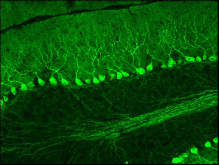
The complementary DNA (cDNA) of a derivative of green
fluorescent protein (GFP) from a jelly fish, was introduced
to the genome encoding G-subtrate by knock-in technique.
G-substrate is specifically localized in cerebellar Purkinje
cells (diameter of 30 m in mice), which are the largest neurons
in the mammalian central nervous system. Cerebellum plays
important roles in motor learning, motor coordination and
non-declarative memories. Cerebellar Purkinje
cells is thought to be a essential neuronal component for these
brain functions. Neuronal plasticity
(flexibility of neuronal transmission) called, long-term
depression, underlies these cerebellar functions.
By expressing GFP, the shape and tracks of Purkinje cells are easily visible under fluorescence microscope. In the picture, the Purkinje cell bodies have strong green fluorescence and a round shape. Purkinje cell dendrites, which enter the molecular layer, are observed as spurs going upward. Also bundles of axons going to cerebellar nuclears are also observed.
The GFP introduced into the specific neurons allows us not only to track the neuronal pathways but also the shape of cells. The GFP gene is often used as a reporter of the expression under a certain promoter or the general expression of the target protein.
Many fluorescent bacteria, fly, yeast, and mammalian cells are developed using GFP. In addition to fluorescent mice, "fluorescent pigs" were generated by Taiwan's group by transgenic technology. If you want to see glow-in-dark FLUORESCENT PIG, Click here.